Leg bone in vertebrates
The
tibia
(
;
pl.
:
tibiae
or
tibias
), also known as the
shinbone
or
shankbone
, is the larger, stronger, and anterior (frontal) of the two
bones
in the
leg
below the
knee
in
vertebrates
(the other being the
fibula
, behind and to the outside of the tibia); it connects the knee with the
ankle
. The tibia is found on the
medial
side of the leg next to the fibula and closer to the
median plane
. The tibia is connected to the fibula by the
interosseous membrane of leg
, forming a type of
fibrous joint
called a
syndesmosis
with very little movement. The tibia is named for the flute
tibia
. It is the second largest bone in the
human body
, after the
femur
. The leg bones are the strongest
long bones
as they support the rest of the body.
Structure
[
edit
]
In
human anatomy
, the tibia is the second largest bone next to the
femur
. As in other vertebrates the tibia is one of two bones in the lower leg, the other being the fibula, and is a component of the knee and ankle joints.
The
ossification
or formation of the bone starts from three centers, one in the shaft and one in each extremity.
The tibia is categorized as a
long bone
and is as such composed of a
diaphysis
and two
epiphyses
. The diaphysis is the midsection of the tibia, also known as the
shaft
or body. While the epiphyses are the two rounded extremities of the bone; an
upper
(also known as superior or proximal) closest to the
thigh
and a
lower
(also known as inferior or distal) closest to the
foot
. The tibia is most contracted in the lower third and the distal extremity is smaller than the proximal.
Upper extremity
[
edit
]
Condyles of tibia
[
edit
]
 Upper surface of right tibia. (Anterior is at top.)
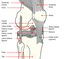 Knee
Knee
The proximal or upper extremity of the tibia is expanded in the transverse plane with a
medial
and
lateral condyle
, which are both flattened in the horizontal plane. The medial condyle is the larger of the two and is better supported over the
shaft
. The upper surfaces of the condyles
articulate
with the femur to form the tibiofemoral joint, the weightbearing part of the kneejoint.
[1]
The medial and lateral condyle are separated by the
intercondylar area
, where the
cruciate ligaments
and the
menisci
attach. Here the
medial
and
lateral intercondylar tubercle
forms the
intercondylar eminence
. Together with the medial and lateral condyle the intercondylar region forms the
tibial plateau
, which both articulates with and is anchored to the
lower extremity of the femur
. The intercondylar eminence divides the intercondylar area into an
anterior
and
posterior part
. The anterolateral region of the anterior intercondylar area are perforated by numerous small openings for
nutrient arteries
.
[1]
The articular surfaces of both condyles are concave, particularly centrally. The flatter outer margins are in contact with the menisci. The medial condyles superior surface is oval in form and extends laterally onto the side of
medial intercondylar tubercle
. The lateral condyles superior surface is more circular in form and its medial edge extends onto the side of the
lateral intercondylar tubercle
. The posterior surface of the medial condyle bears a horizontal groove for part of the attachment of the
semimembranosus muscle
, whereas the lateral condyle has a circular facet for articulation with the
head of the fibula
.
[1]
Beneath the condyles is the
tibial tuberosity
which serves for attachment of the
patellar ligament
, a continuation of the
quadriceps femoris muscle
.
[1]
Facets
[
edit
]
The superior articular surface presents two smooth articular
facets
.
- The
medial facet
, oval in shape, is slightly concave from side to side, and from before backward.
- The
lateral
, nearly circular, is concave from side to side, but slightly convex from before backward, especially at its posterior part, where it is prolonged on to the posterior surface for a short distance.
The central portions of these facets articulate with the condyles of the
femur
, while their peripheral portions support the
menisci
of the
knee
joint, which here intervene between the two bones.
Intercondyloid eminence
[
edit
]
Between the articular facets in the
intercondylar area
, but nearer the posterior than the anterior aspect of the bone, is the
intercondyloid eminence
(
spine of tibia
), surmounted on either side by a prominent tubercle, on to the sides of which the articular facets are prolonged; in front of and behind the intercondyloid eminence are rough depressions for the attachment of the
anterior
and
posterior cruciate ligaments
and the menisci.
Surfaces
[
edit
]
The
anterior surfaces
of the condyles are continuous with one another, forming a large somewhat flattened area; this area is triangular, broad above, and perforated by large vascular foramina; narrow below where it ends in a large oblong elevation, the
tuberosity of the tibia
, which gives attachment to the
patellar ligament
; a
bursa
intervenes between the deep surface of the ligament and the part of the bone immediately above the tuberosity.
Posteriorly,
the condyles are separated from each other by a shallow depression, the
posterior intercondyloid fossa
, which gives attachment to part of the
posterior cruciate ligament
of the
knee-joint
. The medial condyle presents posteriorly a deep transverse groove, for the insertion of the tendon of the
semimembranosus
.
Its
medial surface
is convex, rough, and prominent; it gives attachment to the
medial collateral ligament
.
The lateral condyle presents posteriorly a flat articular facet, nearly circular in form, directed downward, backward, and lateralward, for articulation with the head of the fibula. Its
lateral surface
is convex, rough, and prominent in front: on it is an eminence, situated on a level with the upper border of the tuberosity and at the junction of its anterior and lateral surfaces, for the attachment of the
iliotibial band
. Just below this a part of the
extensor digitorum longus
takes origin and a slip from the tendon of the
biceps femoris
is inserted.
Shaft
[
edit
]
 Bones of the right leg. Anterior surface
The shaft or body of the tibia is triangular in cross-section and forms three borders: an anterior, medial, and lateral or interosseous border. These three borders form three surfaces: the medial, lateral, and posterior.
[2]
The forward flat part of the tibia is called the fibia, often confused with the fibula.
[3]
[
failed verification
]
Borders
[
edit
]
The
anterior crest or border
, the most prominent of the three, commences above at the
tuberosity
, and ends below at the anterior margin of the
medial malleolus
. It is sinuous and prominent in the upper two-thirds of its extent, but smooth and rounded below; it gives attachment to the
deep fascia
of the leg.
The
medial border
is smooth and rounded above and below, but more prominent in the center. It begins at the back part of the medial condyle, and ends at the posterior border of the medial malleolus; its upper part gives attachment to the tibial collateral ligament of the knee-joint to the extent of about 5 cm., and insertion to some fibers of the
popliteus muscle
. From its middle third some fibers of the
soleus
and
flexor digitorum longus muscles
take origin.
The
interosseous crest or lateral border
is thin and prominent, especially its central part, and gives attachment to the
interosseous membrane
; it commences above in front of the fibular articular facet, and bifurcates below, to form the boundaries of a triangular rough surface, for the attachment of the interosseous ligament connecting the tibia and fibula.
Surfaces
[
edit
]
The
medial surface
is smooth, convex, and broader above than below; its upper third, directed forward and medialward, is covered by the
aponeurosis
derived from the tendon of the
sartorius
, and by the tendons of the
Gracilis
and
Semitendinosus
, all of which are inserted nearly as far forward as the anterior crest; in the rest of its extent it is
subcutaneous
.
The
lateral surface
is narrower than the medial; its upper two-thirds present a shallow groove for the origin of the Tibialis anterior; its lower third is smooth, convex, curves gradually forward to the anterior aspect of the bone, and is covered by the tendons of the
Tibialis anterior
,
Extensor hallucis longus
, and
Extensor digitorum longus
, arranged in this order from the medial side.
The
posterior surface
presents, at its upper part, a prominent ridge, the popliteal line, which extends obliquely downward from the back part of the articular facet for the fibula to the medial border, at the junction of its upper and middle thirds; it marks the lower limit of the insertion of the
Popliteus
, serves for the attachment of the fascia covering this muscle, and gives origin to part of the
Soleus
,
Flexor digitorum longus
, and
Tibialis posterior
. The triangular area, above this line, gives insertion to the Popliteus. The middle third of the posterior surface is divided by a vertical ridge into two parts; the ridge begins at the popliteal line and is well-marked above, but indistinct below; the medial and broader portion gives origin to the Flexor digitorum longus, the lateral and narrower to part of the
Tibialis posterior
. The remaining part of the posterior surface is smooth and covered by the Tibialis posterior,
Flexor digitorum longus
, and
Flexor hallucis longus
. Immediately below the popliteal line is the nutrient foramen, which is large and directed obliquely downward.
Lower extremity
[
edit
]
 Lower extremity of right tibia seen from the front
 Lower extremity of right tibia seen from the back
The distal end of the tibia is much smaller than the proximal end and presents five surfaces; it is prolonged downward on its medial side as a strong pyramidal process, the
medial malleolus
. The lower extremity of the tibia together with the fibula and
talus
forms the
ankle joint
.
Surfaces
[
edit
]
The
inferior articular surface
is quadrilateral, and smooth for articulation with the talus. It is concave from before backward, broader in front than behind, and traversed from before backward by a slight elevation, separating two depressions. It is continuous with that on the medial malleolus.
The
anterior surface
of the lower extremity is smooth and rounded above, and covered by the tendons of the Extensor muscles; its lower margin presents a rough transverse depression for the attachment of the articular capsule of the ankle-joint.
The
posterior surface
is traversed by a shallow groove directed obliquely downward and medialward, continuous with a similar groove on the posterior surface of the talus and serving for the passage of the tendon of the
Flexor hallucis longus
.
The
lateral surface
presents a triangular rough depression for the attachment of the inferior interosseous ligament connecting it with the fibula; the lower part of this depression is smooth, covered with cartilage in the fresh state, and articulates with the fibula. The surface is bounded by two prominent borders (the
anterior
and posterior colliculi), continuous above with the
interosseous crest
; they afford attachment to the anterior and posterior ligaments of the lateral malleolus.
The
medial surface
-- see
medial malleolus
for details.
Fractures
[
edit
]
Ankle fractures
of the tibia have several classification systems based on location or mechanism:
Blood supply
[
edit
]
The tibia is supplied with blood from two sources: A
nutrient artery
, as the main source, and
periosteal
vessels derived from the
anterior tibial artery
.
[4]
Joints
[
edit
]
The tibia is a part of four joints; the knee, ankle,
superior
and
inferior tibiofibular joint
.
In the knee the tibia forms one of the two
articulations
with the
femur
, often referred to as the
tibiofemoral components
of the knee joint.;
[5]
[6]
it is the weightbearing part of the knee joint.
[2]
The tibiofibular joints are the articulations between the tibia and fibula which allows very little movement.
[
citation needed
]
The
proximal tibiofibular joint
is a small
plane joint
. The joint is formed between the undersurface of the
lateral tibial condyle
and the
head of fibula
. The
joint capsule
is reinforced by
anterior
and
posterior ligament of the head of the fibula
.
[2]
The
distal tibiofibular joint
(tibiofibular syndesmosis) is formed by the rough, convex surface of the medial side of the distal end of the fibula, and a rough concave surface on the lateral side of the tibia.
[2]
The part of the ankle joint known as the talocrural joint, is a
synovial
hinge joint
that connects the distal ends of the tibia and fibula in the lower limb with the proximal end of the talus. The articulation between the tibia and the talus bears more weight than between the smaller fibula and the talus.
[
citation needed
]
 Plan of ossification of the tibia. From three centers.
 Epiphysial lines of tibia and fibula in a young adult. Anterior aspect.
Development
[
edit
]
The tibia is
ossified
from three
centers
: a
primary center
for the
diaphysis
(shaft) and a secondary center for each
epiphysis
(extremity). Ossification begins in the center of the body, about the seventh week of fetal life, and gradually extends toward the extremities.
The center for the upper epiphysis appears before or shortly after birth at close to 34 weeks gestation; it is flattened in form, and has a thin tongue-shaped process in front, which forms the
tuberosity
; that for the lower epiphysis appears in the second year.
The lower epiphysis fuses with the tibial shaft at about the eighteenth, and the upper one fuses about the twentieth year.
Two additional centers occasionally exist, one for the tongue-shaped process of the upper epiphysis, which forms the tuberosity, and one for the
medial malleolus
.
Function
[
edit
]
Muscle attachments
[
edit
]
Strength
[
edit
]
The tibia has been modeled as taking an axial force during walking that is up to 4.7 bodyweight. Its
bending moment
in the sagittal plane in the late stance phase is up to 71.6 bodyweight times millimetre.
[8]
Clinical significance
[
edit
]
Fracture
[
edit
]
Fractures
of the tibia
can be divided into those that only involve the tibia;
bumper fracture
,
Segond fracture
,
Gosselin fracture
,
toddler's fracture
, and those including both the tibia and
fibula
;
trimalleolar fracture
,
bimalleolar fracture
,
Pott's fracture
.
Society and culture
[
edit
]
In
Judaism
, the tibia, or shankbone, of a goat or sheep is used in the
Passover Seder plate
.
Other animals
[
edit
]
The structure of the tibia in most other
tetrapods
is essentially similar to that in humans. The tuberosity of the tibia, a crest to which the
patellar ligament
attaches in mammals, is instead the point for the tendon of the
quadriceps
muscle in reptiles, birds, and amphibians, which have no
patella
.
[9]
Additional images
[
edit
]
-
Shape of right tibia
-
3D image
-
Longitudinal section of tibia showing interior
-
Right knee-joint. Anterior view.
-
Right knee joint from the front, showing interior ligaments
-
Left knee joint from behind, showing interior ligaments
-
Left talocrural joint
-
Coronal section through right talocrural and talocalcaneal joints
-
Dorsum of Foot. Ankle joint. Deep dissection
-
Dorsum of Foot. Ankle joint. Deep dissection
-
Ankle joint. Deep dissection. Anterior view
-
Bones of the right leg. Anterior surface
-
Bones of the right leg. Posterior surface
-
Dorsum of Foot. Ankle joint. Deep dissection.
-
Ankle joint. Deep dissection.
-
Ankle joint. Deep dissection.
-
Ankle joint. Deep dissection.
-
Ankle joint. Deep dissection.
-
Tibia Anatomy
See also
[
edit
]
References
[
edit
]
 This article incorporates text in the
public domain
from
page 256
of the 20th edition of
Gray's Anatomy
(1918)
This article incorporates text in the
public domain
from
page 256
of the 20th edition of
Gray's Anatomy
(1918)
- ^
a
b
c
d
Drake, Richard L.; Vogl, A. Wayne; Mitchell, Adam W. M. (2010).
Gray´s Anatomy for Students
(2nd ed.). Churchill Livingstone/Elsevier. pp. 558?560.
ISBN
978-0-443-06952-9
.
[
page needed
]
- ^
a
b
c
d
Drake, Richard L.; Vogl, A. Wayne; Mitchell, Adam W. M. (2010).
Gray´s Anatomy for Students
(2nd ed.). Churchill Livingstone/Elsevier. pp. 584?588.
ISBN
978-0-443-06952-9
.
- ^
"Chapter 12: THE BONES OF THE LOWER LIMB"
.
www.dartmouth.edu
. Retrieved
2018-06-17
.
- ^
Nelson G, Kelly P, Peterson L, Janes J (1960). "Blood supply of the human tibia".
J Bone Joint Surg Am
.
42-A
(4): 625?36.
doi
:
10.2106/00004623-196042040-00007
.
PMID
13854090
.
- ^
Rytter S, Egund N, Jensen LK, Bonde JP (2009).
"Occupational kneeling and radiographic tibiofemoral and patellofemoral osteoarthritis"
.
J Occup Med Toxicol
.
4
(1): 19.
doi
:
10.1186/1745-6673-4-19
.
PMC
2726153
.
PMID
19594940
.
- ^
Gill TJ, Van de Velde SK, Wing DW, Oh LS, Hosseini A, Li G (December 2009).
"Tibiofemoral and patellofemoral kinematics after reconstruction of an isolated posterior cruciate ligament injury: in vivo analysis during lunge"
.
Am J Sports Med
.
37
(12): 2377?85.
doi
:
10.1177/0363546509341829
.
PMC
3832057
.
PMID
19726621
.
- ^
Bojsen-Møller, Finn; Simonsen, Erik B.; Tranum-Jensen, Jørgen (2001).
Bevægeapparatets anatomi
[
Anatomy of the Locomotive Apparatus
] (in Danish) (12th ed.). Munksgaard Danmark. pp. 364?367.
ISBN
978-87-628-0307-7
.
- ^
Wehner, T; Claes, L; Simon, U (2009). "Internal loads in the human tibia during gait".
Clin Biomech
.
24
(3): 299?302.
doi
:
10.1016/j.clinbiomech.2008.12.007
.
PMID
19185959
.
- ^
Romer, Alfred Sherwood; Parsons, Thomas S. (1977).
The Vertebrate Body
. Philadelphia, PA: Holt-Saunders International. p. 205.
ISBN
0-03-910284-X
.