Eye that appears red due to illness or injury
Medical condition
| Red eye
|
|---|
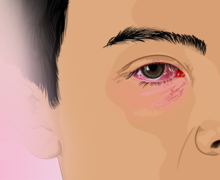 |
| Subconjunctival hemorrhage causing red coloration as result of ruptured blood vessel in the eye
|
| Specialty
| Ophthalmology
 |
|---|
A
red eye
is an
eye
that appears
red
due to
illness
or
injury
. It is usually
injection and prominence
of the superficial
blood vessels
of the
conjunctiva
, which may be caused by disorders of these or adjacent structures.
Conjunctivitis
and
subconjunctival hemorrhage
are two of the less serious but more common causes.
Management includes assessing whether emergency action (including referral) is needed, or whether treatment can be accomplished without additional resources.
Slit lamp examination
is invaluable in diagnosis but initial assessment can be performed using a careful history, testing vision (
visual acuity
), and carrying out a
penlight examination
.
Diagnosis
[
edit
]
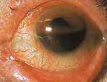 Hyphaema
? showing blood filling the anterior chamber, causing a horizontal fluid level
Hyphaema
? showing blood filling the anterior chamber, causing a horizontal fluid level
Particular
signs
and
symptoms
may indicate that the cause is serious and requires immediate attention.
[1]
Seven such signs are:
The most useful is a smaller pupil in the red eye than the non-red eye (opposite eye) and sensitivity to bright light.
[2]
Reduced visual acuity
[
edit
]
A reduction in
visual acuity
in a 'red eye' is indicative of serious ocular disease,
[3]
such as
keratitis
,
iridocyclitis
, and
glaucoma
, and never occurs in simple
conjunctivitis
without accompanying corneal involvement.
Ciliary flush
[
edit
]
Ciliary flush is usually present in eyes with corneal inflammation, iridocyclitis or
acute glaucoma
, though not simple conjunctivitis.
A ciliary flush is a ring of
red
or
violet
spreading out from around the
cornea
of the eye.
Corneal abnormalities
[
edit
]
The
cornea
is required to be transparent to transmit light to the retina. Because of injury, infection or inflammation, an area of opacity may develop which can be seen with a penlight or
slit lamp
. In rare instances, this opacity is congenital.
[4]
In some, there is a family history of corneal growth disorders which may be progressive with age. Much more commonly, misuse of contact lenses may be a precipitating factor. Whichever, it is always potentially serious and sometimes necessitates urgent treatment and
corneal opacities
are the fourth leading cause of blindness.
Opacities may be keratic, that is, due to the deposition of inflammatory cells, hazy, usually from corneal
edema
, or they may be localized in the case of
corneal ulcer
or
keratitis
.
Corneal epithelial disruptions may be detected with
fluorescein
staining of the eye, and careful observation with
cobalt
-
blue
light
.
Corneal epithelial disruptions would stain
green
, which represents some injury of the corneal epithelium.
These types of disruptions may be due to
corneal inflammations
or
physical trauma
to the cornea, such as a foreign body.
Pupillary abnormalities
[
edit
]
In an eye with iridocyclitis, (inflammation of both the iris and ciliary body), the involved
pupil
will be smaller than the uninvolved, due to reflex
muscle spasm
of the
iris sphincter muscle
.
Generally, conjunctivitis does not affect the pupils.
With
acute angle-closure glaucoma
, the pupil is generally fixed in mid-position, oval, and responds sluggishly to light, if at all.
Shallow anterior chamber depth may indicate a predisposition to one form of glaucoma (narrow angle) but requires
slit-lamp examination
or other special techniques to determine it.
In the presence of a "red eye", a shallow anterior chamber may indicate acute
glaucoma
, which requires immediate attention.
Abnormal intraocular pressure
[
edit
]
Intraocular pressure
should be measured as part of a routine
eye examination
.
It is usually only elevated by iridocyclitis or acute-closure glaucoma, but not by relatively benign conditions.
In iritis and traumatic perforating ocular injuries, the intraocular pressure is usually low.
Severe pain
[
edit
]
Those with
conjunctivitis
may report mild irritation or scratchiness, but never extreme pain, which is an indicator of more serious disease such as
keratitis
,
corneal ulceration
,
iridocyclitis
, or acute
glaucoma
.
Differential diagnosis
[
edit
]
Of the many causes,
conjunctivitis
is the most common.
[1]
Others include:
Usually nonurgent
[
edit
]
- airborne
eye irritants
- blepharitis
[5]
? a usually chronic inflammation of the eyelids with scaling, sometimes resolving spontaneously
- drugs:
medications
or
recreational drug use
- dry eye syndrome
? caused by either decreased tear production or increased tear film evaporation which may lead to irritation and redness
[8]
 Acute glaucoma, angle closure type
- subconjunctival hemorrhage
[1]
? a sometimes dramatic, but usually harmless, bleeding underneath the conjunctiva most often from spontaneous rupture of the small, fragile blood vessels, commonly from a cough or sneeze
- inflamed
pterygium
[9]
? a benign, triangular, horizontal growth of the conjunctiva, arising from the inner side, at the level of contact of the upper and lower eyelids, associated with exposure to sunlight, low humidity and dust. It may be more common in occupations such as farming and welding.
- inflamed
pinguecula
[10]
? a yellow-white deposit close to the junction between the cornea and sclera, on the conjunctiva. It is most prevalent in tropical climates with much UV exposure. Although harmless, it can occasionally become inflamed.
- tiredness
- episcleritis
[11]
? most often a mild, inflammatory disorder of the 'white' of the eye unassociated with eye complications in contrast to scleritis, and responding to topical medications such as anti-inflammatory drops.
Usually urgent
[
edit
]
- acute closed-angle glaucoma
[12]
? implies injury to the optic nerve with the potential for irreversible vision loss which may be permanent unless treated quickly, as a result of increased pressure within the eyeball. Not all forms of glaucoma are acute, and not all are associated with increased intraocular pressure.
- eye injury
- keratitis
[12]
? a potentially serious inflammation or injury to the cornea (window), often associated with significant pain, light intolerance, and deterioration in vision. Numerous causes include virus infection. Injury from contact lenses can lead to keratitis.
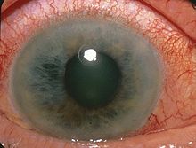
 Eye with iritis showing ciliary flush
- iritis
[1]
? together with the
ciliary body
and
choroid
, the iris makes up the
uvea
, part of the middle, pigmented, structures of the eye. Inflammation of this layer (uveitis) requires urgent control and is estimated to be responsible for 10% of blindness in the United States.
- scleritis
[13]
? a serious inflammatory condition, often painful, that can result in permanent vision loss, and without an identifiable cause in half of those presenting with it. About 30?40% have an underlying systemic
autoimmune
condition.
- tick-borne illnesses like
Rocky Mountain spotted fever
[14]
? the eye is not primarily involved, but the presence of conjunctivitis, along with fever and rash, may help with the diagnosis in appropriate circumstances.
See also
[
edit
]
References
[
edit
]
- ^
a
b
c
d
Cronau, H; Kankanala, RR; Mauger, T (Jan 15, 2010). "Diagnosis and management of red eye in primary care".
American Family Physician
.
81
(2): 137?44.
PMID
20082509
.
- ^
Narayana, S; McGee, S (November 2015).
"Bedside Diagnosis of the 'Red Eye': A Systematic Review"
.
The American Journal of Medicine
.
128
(11): 1220?1224.e1.
doi
:
10.1016/j.amjmed.2015.06.026
.
PMID
26169885
.
- ^
Leibowitz HM (2000). "The red eye".
N Engl J Med
.
343
(5): 345?51.
doi
:
10.1056/nejm200008033430507
.
PMID
10922425
.
- ^
Rezende RA, Uchoa UB, Uchoa R, Rapuano CJ, Laibson PR, Cohen EJ (2004). "Congenital corneal opacities in a cornea referral practice".
Cornea
.
23
(6): 565?70.
doi
:
10.1097/01.ico.0000126317.90271.d8
.
PMID
15256994
.
S2CID
9031282
.
- ^
Jackson WB (April 2008). "Blepharitis: current strategies for diagnosis and management".
Can J Ophthalmol
.
43
(2): 170?79.
doi
:
10.3129/i08-016
.
PMID
18347619
.
- ^
Yazulla, S (September 2008).
"Endocannabinoids in the retina: from marijuana to neuroprotection"
.
Progress in Retinal and Eye Research
.
27
(5): 501?26.
doi
:
10.1016/j.preteyeres.2008.07.002
.
PMC
2584875
.
PMID
18725316
.
- ^
American Psychiatric Association. Diagnostic and Statistical Manual of Mental Disorders, Fourth Edition, Text Revision (DSM-IV-TR). Washington DC: American Psychiatric Association; 2000.
- ^
"Keratoconjunctivitis, Sicca"
.
eMedicine
.
WebMD, Inc.
January 27, 2010
. Retrieved
September 3,
2010
.
- ^
Bradley JC, Yang W, Bradley RH, Reid TW, Schwab IR (July 2010). "The science of pterygia".
Br J Ophthalmol
.
94
(7): 815?20.
doi
:
10.1136/bjo.2008.151852
.
PMID
19515643
.
S2CID
15507689
.
- ^
Sutphin, John, ed. 2007?2008 Basic and Clinical Science Course Section 8: External Disease and Cornea. American Academy Ophthalmology. p. 365.
ISBN
1-56055-814-8
.
- ^
Jabs DA, Mudun A, Dunn JP, Marsh MJ (October 2000). "Episcleritis and scleritis: clinical features and treatment results".
Am J Ophthalmol
.
130
(4): 469?76.
doi
:
10.1016/S0002-9394(00)00710-8
.
PMID
11024419
.
- ^
a
b
Dargin JM, Lowenstein RA (February 2008). "The painful eye".
Emerg Med Clin North Am
.
26
(1): 199?216.
doi
:
10.1016/j.emc.2007.10.001
.
PMID
18249263
.
- ^
Sims, J (December 2012).
"Scleritis: presentations, disease associations and management"
.
Postgrad Med J
.
88
(1046): 713?18.
doi
:
10.1136/postgradmedj-2011-130282
.
PMID
22977282
.
S2CID
37084152
.
- ^
https://www.cdc.gov/mmwr/pdf/rr/rr5504.pdf
[
bare URL PDF
]
External links
[
edit
]