Flat bone in the middle front part of the rib cage
The
sternum
(
pl.
:
sternums
or
sterna
) or
breastbone
is a long
flat bone
located in the central part of the
chest
. It connects to the
ribs
via
cartilage
and forms the front of the
rib cage
, thus helping to protect the
heart
,
lungs
, and major
blood vessels
from injury. Shaped roughly like a
necktie
, it is one of the largest and longest flat bones of the body. Its three regions are the manubrium, the body, and the
xiphoid process
.
[1]
The word
sternum
originates from Ancient Greek στ?ρνον (
sternon
) 'chest'.
Structure
[
edit
]
The sternum is a narrow,
flat bone
, forming the middle portion of the front of the
chest
. The top of the sternum supports the
clavicles
(collarbones) and its edges join with the
costal cartilages
of the first two pairs of
ribs
. The inner surface of the sternum is also the attachment of the
sternopericardial ligaments
.
[2]
Its top is also connected to the
sternocleidomastoid muscle
. The sternum consists of three main parts, listed from the top:
In its natural position, the sternum is angled obliquely, downward and forward. It is slightly convex in front and concave behind; broad above, shaped like a "T", becoming narrowed at the point where the manubrium joins the body, after which it again widens a little to below the middle of the body, and then narrows to its lower extremity. In adults the sternum is on average about 1.7 cm longer in the male than in the female.
[
citation needed
]
Manubrium
[
edit
]
 Shape of manubrium
The manubrium (
Latin
for 'handle') is the broad upper part of the sternum. It has a
quadrangular
shape, narrowing from the top, which gives it four borders. The
suprasternal notch
(jugular notch) is located in the middle at the upper broadest part of the manubrium. This notch can be felt between the two
clavicles
. On either side of this notch are the right and left
clavicular notches
.
[1]
The manubrium joins with the body of the sternum, the clavicles and the cartilages of the first pair of
ribs
. The inferior border, oval and rough, is covered with a thin layer of cartilage for articulation with the body. The lateral borders are each marked above by a depression for the first
costal cartilage
, and below by a small facet, which, with a similar facet on the upper angle of the body, forms a notch for the reception of the costal cartilage of the second rib. Between the depression for the first costal cartilage and the demi-facet for the second is a narrow, curved edge, which slopes from above downward towards the middle. Also, the superior sternopericardial ligament attaches the
pericardium
to the posterior side of the manubrium.
Body
[
edit
]
 3D illustration of the body of sternum.
The body, or gladiolus, is the longest sternal part. It is flat and considered to have only a front and back surface. It is flat on the front, directed upward and forward, and marked by three transverse ridges which cross the bone opposite the third, fourth, and fifth articular depressions. The
pectoralis major
attaches to it on either side. At the junction of the third and fourth parts of the body is occasionally seen an orifice, the sternal foramen, of varying size and form. The posterior surface, slightly concave, is also marked by three transverse lines, less distinct, however, than those in front; from its lower part, on either side, the
transversus thoracis
takes origin.
The
sternal angle
is located at the point where the body joins the manubrium. The sternal angle can be felt at the point where the sternum projects farthest forward. However, in some people the sternal angle is concave or rounded. During physical examinations, the sternal angle is a useful landmark because the second rib attaches here.
[1]
Each outer border, at its superior angle, has a small facet, which with a similar facet on the manubrium, forms a cavity for the cartilage of the second rib; below this are four angular depressions which receive the cartilages of the third, fourth, fifth, and sixth ribs. The inferior angle has a small facet, which, with a corresponding one on the xiphoid process, forms a notch for the cartilage of the seventh rib. These articular depressions are separated by a series of curved interarticular intervals, which diminish in length from above downward, and correspond to the intercostal spaces. Most of the
cartilages
belonging to the
true ribs
, articulate with the sternum at the lines of junction of its primitive component segments. This is well seen in some other vertebrates, where the parts of the bone remain separated for longer.
[
citation needed
]
The upper border is oval and articulates with the manubrium, at the sternal angle. The lower border is narrow, and articulates with the
xiphoid process
.
Xiphoid process
[
edit
]
 3D illustration of the xiphoid process.
Located at the inferior end of the sternum is the pointed
xiphoid process
. Improperly performed chest compressions during
cardiopulmonary resuscitation
can cause the xiphoid process to snap off, driving it into the liver which can cause a fatal hemorrhage.
[1]
The sternum is composed of highly
vascular
tissue, covered by a thin layer of compact bone which is thickest in the manubrium between the
articular facets
for the
clavicles
. The inferior sternopericardial ligament attaches the pericardium to the posterior xiphoid process.
Joints
[
edit
]
The cartilages of the top five ribs join with the sternum at the
sternocostal joints
. The right and left clavicular notches articulate with the right and left clavicles, respectively. The costal cartilage of the second rib articulates with the sternum at the sternal angle making it easy to locate.
[3]
The
transversus thoracis
muscle is innervated by one of the
intercostal nerves
and superiorly attaches at the posterior surface of the lower sternum. Its inferior attachment is the internal surface of costal cartilages two through six and works to depress the ribs.
[4]
Development
[
edit
]
 Figure 4
Ossification
Figure 4
Ossification
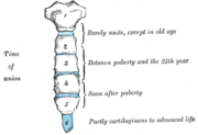 Figure 5
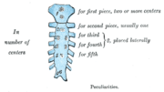 Figure 6
Peculiarities
Figure 6
Peculiarities
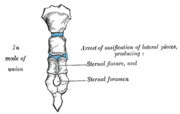 Figure 7
The sternum develops from two cartilaginous bars one on the left and one on the right, connected with the cartilages of the ribs on each side.
[5]
These two bars fuse together along the middle to form the cartilaginous sternum which is ossified from six centers: one for the manubrium, four for the body, and one for the
xiphoid process
.
The
ossification
centers appear in the intervals between the articular depressions for the
costal cartilages
, in the following order: in the manubrium and first piece of the body, during the sixth month of fetal life; in the second and third pieces of the body, during the seventh month of fetal life; in its fourth piece, during the first year after birth; and in the xiphoid process, between the fifth and eighteenth years.
The centers make their appearance at the upper parts of the segments, and proceed gradually downward. To these may be added the occasional existence of two small
episternal
centers, which make their appearance one on either side of the
jugular notch
; they are probably vestiges of the episternal bone of the monotremata and lizards.
[
citation needed
]
Occasionally some of the segments are formed from more than one center, the number and position of which vary [Fig. 6]. Thus, the first piece may have two, three, or even six centers.
When two are present, they are generally situated one above the other, the upper being the larger; the second piece has seldom more than one; the third, fourth, and fifth pieces are often formed from two centers placed laterally, the irregular union of which explains the rare occurrence of the sternal foramen [Fig. 7], or of the vertical fissure which occasionally intersects this part of the bone constituting the malformation known as
fissura sterni;
these conditions are further explained by the manner in which the cartilaginous sternum is formed.
More rarely still the upper end of the sternum may be divided by a
fissure
. Union of the various centers of the body begins about
puberty
, and proceeds from below upward [Fig. 5]; by the age of 25 they are all united.
The xiphoid process may become joined to the body before the age of thirty, but this occurs more frequently after forty; on the other hand, it sometimes remains ununited in old age. In advanced life the manubrium is occasionally joined to the body by bone. When this takes place, however, the bony tissue is generally only superficial, the central portion of the intervening cartilage remaining unossified.
The body of the sternum is formed by the fusion of four segments called
sternebrae
.
[6]
Variations
[
edit
]
In 2.5?13.5% of the population, a foramen known as
sternal foramen
may be presented at the lower third of the sternal body.
[7]
In extremely rare cases, multiple foramina may be observed. Fusion of the manubriosternal joint also occurs in around 5% of the population.
[8]
Small ossicles known as
episternal ossicles
may also be present posterior to the superior end of the manubrium.
[9]
Another variant called suprasternal tubercle is formed when the episternal ossicles fuse with the manubrium.
[10]
Clinical significance
[
edit
]
Bone marrow biopsy
[
edit
]
Because the sternum contains
bone marrow
, it is sometimes used as a site for
bone marrow biopsy
. In particular, patients with a high
BMI
(obese or grossly overweight) may present with excess tissue that makes access to traditional marrow biopsy sites such as the
pelvis
difficult.
Sternal opening
[
edit
]
A somewhat rare
congenital disorder
of the sternum sometimes referred to as an
anatomical variation
is a sternal foramen, a single round hole in the sternum that is present from birth and usually is off-centered to the right or left, commonly forming in the 2nd, 3rd, and 4th segments of the breastbone body. Congenital sternal foramina can often be mistaken for bullet holes.
[11]
They are usually
without symptoms
but can be problematic if acupuncture in the area is intended.
[12]
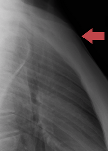 Manubrium sternal dislocation
Fractures
[
edit
]
Fractures of the sternum
are rather uncommon. They may result from trauma, such as when a driver's chest is forced into the
steering column
of a
car
in a
car accident
. A fracture of the sternum is usually a
comminuted
fracture. The most common site of sternal fractures is at the
sternal angle
. Some studies reveal that repeated punches or continual beatings, sometimes called "breastbone punches", to the sternum area have also caused fractured sternums. Those are known to have occurred in contact sports such as hockey and football. Sternal fractures are frequently associated with underlying injuries such as
pulmonary contusions
, or bruised lung tissue.
[13]
Dislocation
[
edit
]
A manubriosternal dislocation is rare and usually caused by severe trauma. It may also result from minor trauma where there is a precondition of arthritis.
[14]
Sternotomy
[
edit
]
The breastbone is sometimes cut open (a
median sternotomy
) to gain access to the thoracic contents when performing
cardiothoracic surgery
. Surgical fixation of sternotomy is achieved through the use of either wire cerclage or a plate and screw technique. The incidence of sternotomy complications falls within the narrow range of 0.5% to 5%. Nevertheless, these complications can have severe consequences, including increased mortality rates, the need for reoperation, and a mortality rate as high as 40%. Such complications often entail issues like dehiscence and sternal non-union, primarily stemming from lateral forces exerted during post-operative activities such as coughing and sneezing.
Resection
[
edit
]
The sternum can be totally removed (resected) as part of a radical surgery, usually to surgically treat a malignancy, either with or without a mediastinal
lymphadenectomy
(
Current Procedural Terminology
codes # 21632 and # 21630, respectively).
Bifid sternum or sternal cleft
[
edit
]
A bifid sternum is an extremely rare congenital abnormality caused by the fusion failure of the sternum.
[15]
This condition results in
sternal cleft
which can be observed at birth without any symptom.
[15]
Other animals
[
edit
]
The sternum, in vertebrate anatomy, is a
flat bone
that lies in the middle front part of the
rib cage
. It is
endochondral
in origin.
[16]
It probably first evolved in early
tetrapods
as an extension of the
pectoral girdle
; it is not found in
fish
. In
amphibians
and
reptiles
, it is typically a shield-shaped structure, often composed entirely of
cartilage
. It is absent in both
turtles
and
snakes
. In
birds
, it is a relatively large bone and typically bears an enormous projecting
keel
to which the flight muscles are attached.
[17]
Only in
mammals
does the sternum take on the elongated, segmented form seen in humans.
Arthropods
[
edit
]
In arthropods, a sternum is the
ventral
part of a segment of
thorax
or
abdomen
.
Etymology
[
edit
]
English
sternum
is a translation of
Ancient Greek
στ?ρνον
,
sternon
.
[18]
The Greek writer
Homer
used the term
στ?ρνον
to refer to the
male chest
,
[19]
[20]
and the term
στ?θο?
,
stithos
to refer to the
chest of both sexes
.
[19]
[20]
The Greek
physician
Hippocrates
used στ?ρνον to refer to the
chest
,
[19]
[20]
and στ?θο? to the
breastbone
.
[19]
[20]
The Greek physician
Galen
was the first to use
στ?ρνον
in the present meaning of
breastbone
.
[19]
[20]
The sternum as the solid bony part of the chest
[21]
can be related to Ancient Greek
στερε??/στερρ??
,
(
stere?s/sterr?s
)
,
[21]
meaning
firm
or
solid
.
[20]
The English term breastbone is actually more like the Latin
os pectoris
,
[22]
[23]
derived from classical Latin
os
, bone
[24]
and
pectus
, chest or breast.
[24]
Confusingly,
pectus
is also used in classical Latin as
breastbone
.
[24]
Additional images
[
edit
]
-
Position of sternum (shown in red). Animation.
-
Sternum seen posteriorly
-
Sternum cut along the frontal plane showing interior of the bone
-
Sternum, lateral aspect
-
Position of the sternum the thoracic cage
-
Computer-generated image of ribcage turntable highlighting the sternum
-
Sternum anatomy
See also
[
edit
]
References
[
edit
]
- ^
a
b
c
d
Saladin, Kenneth S. (2010).
Anatomy and Physiology: The Unity of Form and Function, Fifth Edition
. New York, NY: McGraw-Hill. p.
266
.
ISBN
978-0-07-352569-3
.
- ^
Dyce, Keith M.; Sack, Wolfgang O.; Wensing, C. J. G. (2009-12-03).
Textbook of Veterinary Anatomy
. Elsevier Health Sciences.
ISBN
978-1437708752
.
- ^
Agur, Anne M.R.; Dalley, Arthur F. II (2009).
Grant's Atlas of Anatomy, Twelfth Edition
. Philadelphia, PA: Lippincott Williams and Wilkins. p.
10
.
ISBN
978-0-7817-7055-2
.
- ^
Agur, Anne M.R.; Dalley, Arthur F. II (2009).
Grant's Atlas of Anatomy, Twelfth Edition
. Philadelphia, PA: Lippincott Williams and Wilkins. p.
21
.
ISBN
978-0-7817-7055-2
.
- ^
 This article incorporates
text
available under the
CC BY 4.0
license.
Betts, J Gordon; Desaix, Peter; Johnson, Eddie; Johnson, Jody E; Korol, Oksana; Kruse, Dean; Poe, Brandon; Wise, James; Womble, Mark D; Young, Kelly A (May 14, 2023).
Anatomy & Physiology
. Houston: OpenStax CNX. 7.5 Embryonic development of the axial skeleton.
ISBN
978-1-947172-04-3
.
This article incorporates
text
available under the
CC BY 4.0
license.
Betts, J Gordon; Desaix, Peter; Johnson, Eddie; Johnson, Jody E; Korol, Oksana; Kruse, Dean; Poe, Brandon; Wise, James; Womble, Mark D; Young, Kelly A (May 14, 2023).
Anatomy & Physiology
. Houston: OpenStax CNX. 7.5 Embryonic development of the axial skeleton.
ISBN
978-1-947172-04-3
.
- ^
Clinical Anatomy of the Spine, Spinal Cord, and Ans
. Elsevier. 2014. p. 226.
doi
:
10.1016/c2009-0-42801-0
.
ISBN
978-0-323-07954-9
.
- ^
Choi, Paul J; Iwanaga, Joe; Tubbs, R. Shane (2017).
"A Comprehensive Review of the Sternal Foramina and its Clinical Significance"
.
Cureus
.
9
(12): e1929.
doi
:
10.7759/cureus.1929
.
ISSN
2168-8184
.
PMC
5805319
.
PMID
29456905
.
- ^
Sebes, Ji; Salazar, Je (1983-01-01).
"The manubriosternal joint in rheumatoid disease"
.
American Journal of Roentgenology
.
140
(1): 117?121.
doi
:
10.2214/ajr.140.1.117
.
ISSN
0361-803X
.
PMID
6600299
.
- ^
Stark, P.; Watkins, G. E.; Hildebrandt-Stark, H. E.; Dunbar, R. D. (1987).
"Episternal ossicles"
.
Radiology
.
165
(1): 143?144.
doi
:
10.1148/radiology.165.1.3628759
.
ISSN
0033-8419
.
PMID
3628759
.
- ^
Duraikannu, Chary; Noronha, Olma V; Sundarrajan, Pushparajan (2016).
"MDCT evaluation of sternal variations: Pictorial essay"
.
The Indian Journal of Radiology & Imaging
.
26
(2): 185?194.
doi
:
10.4103/0971-3026.184407
.
ISSN
0971-3026
.
PMC
4931775
.
PMID
27413263
.
- ^
Byers, S.N. (2008).
Introduction to Forensic Anthropology
. Toronto: Pearson.
- ^
Fokin, AA (May 2000). "Cleft sternum and sternal foramen".
Chest Surgery Clinics of North America
.
10
(2): 261?76.
PMID
10803333
.
- ^
Sattler S, Maier RV (2002).
"Pulmonary contusion"
. In Karmy-Jones R, Nathens A, Stern EJ (eds.).
Thoracic Trauma and Critical Care
. Berlin: Springer. pp. 235?243.
ISBN
1-4020-7215-5
. Retrieved
2008-04-21
.
- ^
El Ibrahimi, Abdelhalim; Sbai, Hicham; Kanjaa, Nabil; Shimi, Mohammed; Lakranbi, Marouane; Daoudi, Abdelkrim; Elmrini, Abdelmajid; Smahi, Mohammed (2011).
"Traumatic manubriosternal dislocation: A new method of stabilization postreduction"
.
Journal of Emergencies, Trauma, and Shock
.
4
(2): 317?319.
doi
:
10.4103/0974-2700.82237
.
PMC
3132377
.
PMID
21769224
.
- ^
a
b
Das, Sibes Kumar; Jana, Pulak Kumar; Bairagya, Tapan Das; Ghoshal, Bhaswati (2012-01-01).
"Bifid sternum"
.
Lung India
.
29
(1): 73?75.
doi
:
10.4103/0970-2113.92370
.
ISSN
0970-2113
.
PMC
3276042
.
PMID
22345921
.
- ^
Kardong, Kenneth V. (1995).
Vertebrates: comparative anatomy, function, evolution
. McGraw-Hill. pp. 55, 57.
ISBN
0-697-21991-7
.
- ^
Romer, Alfred Sherwood; Parsons, Thomas S. (1977).
The Vertebrate Body
. Philadelphia, PA: Holt-Saunders International. p. 188.
ISBN
0-03-910284-X
.
- ^
Triepel, H. (1910).
Die anatomischen Namen. Ihre Ableitung und Aussprache. Mit einem Anhang: Biographische Notizen.
(Dritte Auflage). Wiesbaden: Verlag J.F. Bergmann.
- ^
a
b
c
d
e
Hyrtl, J. (1880).
Onomatologia Anatomica. Geschichte und Kritik der anatomischen Sprache der Gegenwart.
Wien: Wilhelm Braumuller. K.K. Hof- und Universitatsbuchhandler.
- ^
a
b
c
d
e
f
Liddell, H.G. & Scott, R. (1940).
A Greek-English Lexicon. revised and augmented throughout by Sir Henry Stuart Jones. with the assistance of. Roderick McKenzie.
Oxford: Clarendon Press.
- ^
a
b
Kraus, L.A. (1844).
Kritisch-etymologisches medicinisches Lexikon
(Dritte Auflage). Gottingen: Verlag der Deuerlich- und Dieterichschen Buchhandlung.
- ^
Schreger, C.H.Th. (1805).
Synonymia anatomica. Synonymik der anatomischen Nomenclatur.
Furth: im Bureau fur Literatur.
- ^
Siebenhaar, F.J. (1850).
Terminologisches Worterbuch der medicinischen Wissenschaften.
(Zweite Auflage). Leipzig: Arnoldische Buchhandlung.
- ^
a
b
c
Lewis, C.T. & Short, C. (1879).
A Latin dictionary founded on Andrews' edition of Freund's Latin dictionary.
Oxford: Clarendon Press.
External links
[
edit
]
 Media related to
Sternum
at Wikimedia Commons
Media related to
Sternum
at Wikimedia Commons